Being called a "glaucoma suspect" does not necessarily mean you have glaucoma. Ocular hypertension does not automatically mean glaucoma either.
These terms describe different combinations of eye pressure, optic-nerve appearance, imaging, visual-field results, drainage-angle anatomy, and risk. The distinction matters because some people need treatment, some need closer testing, and others can be monitored without starting medication immediately.
The three terms are not interchangeable
Ocular hypertension
Ocular hypertension generally means the measured pressure inside the eye is higher than expected, but there is no confirmed glaucomatous optic-nerve damage or matching visual-field loss.
Pressure is a risk factor, not the entire diagnosis. Some eyes tolerate a certain pressure without measurable damage, while others develop glaucoma at pressures that fall within a commonly quoted range.
Glaucoma suspect
A glaucoma suspect has one or more findings that raise concern for future or early glaucoma. That may include:
- Consistently elevated eye pressure.
- A suspicious optic-nerve shape or cup-to-disc ratio.
- Possible thinning on OCT imaging.
- A visual-field result that may fit glaucoma but is not yet confirmed.
- A strong combination of risk factors.
The 2026 American Academy of Ophthalmology Preferred Practice Pattern describes a suspect as someone with elevated pressure, suspicious optic-nerve or retinal nerve-fiber findings, or a suspicious visual field without enough evidence to establish primary open-angle glaucoma.
Glaucoma
Glaucoma is a group of diseases that damage the optic nerve. In primary open-angle glaucoma, diagnosis usually depends on a pattern of structural damage, functional loss, progression, or a convincing combination of findings rather than one isolated pressure reading.
Once nerve tissue and visual function are lost, treatment aims to preserve what remains. That is why careful monitoring before obvious symptoms develop is important.
Why one eye-pressure reading is not enough
Eye pressure varies during the day and can be affected by corneal properties, measurement technique, medications, recent activity, and other factors.
A pressure number should be interpreted with:
- Prior measurements and time of day.
- Central corneal thickness.
- Corneal biomechanics when available.
- Optic-nerve appearance.
- OCT and visual-field trends.
- Drainage-angle anatomy.
- Family and medical history.
A thick cornea can make applanation pressure read higher than the pressure's true effect suggests, while a thin cornea can be associated with underestimated readings and greater glaucoma risk. Pachymetry does not simply "correct" the pressure with one universal formula; it adds context to the full risk assessment.
What each glaucoma test contributes
Tonometry: the pressure measurement
Tonometry measures intraocular pressure. Repeated readings help show whether pressure is consistently elevated, fluctuating, or responding to treatment.
Pachymetry: corneal thickness
Pachymetry measures central corneal thickness. It helps the clinician interpret pressure and risk, but it does not diagnose glaucoma by itself.
Gonioscopy: the drainage angle
Gonioscopy examines the angle where fluid leaves the eye. It distinguishes open-angle anatomy from narrow or closed angles and can reveal pigment, trauma, abnormal vessels, or other secondary causes.
Optic-nerve examination and photography
The clinician evaluates the nerve's rim, cup, symmetry, hemorrhages, and change over time. Baseline photography can make future comparison more reliable.
OCT and visual-field testing
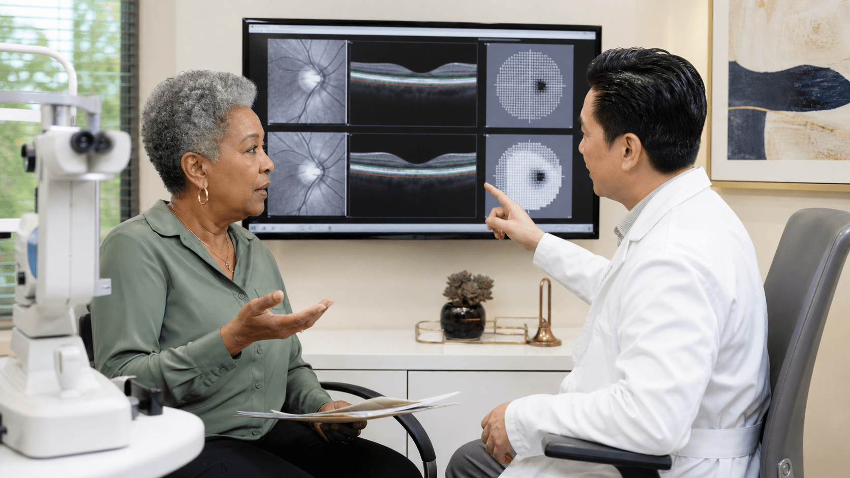
OCT images optic-nerve structures while a visual-field test checks functional vision. Both can inform whether a patient is a suspect or has confirmed damage, but neither printout is a diagnosis alone. Our OCT versus visual-field guide explains what each test measures, why results sometimes disagree, and what to ask at follow-up.
Risk factors that change the follow-up plan
The AAO guideline identifies factors associated with higher risk of developing primary open-angle glaucoma, including:
- Higher intraocular pressure.
- Older age.
- Family history of glaucoma.
- Black or Latino/Hispanic background.
- Thin central cornea.
- Myopia.
- Diabetes and certain vascular factors.
- Optic-disc hemorrhage or more suspicious nerve and field measurements.
Risk is cumulative. The same pressure can lead to different recommendations in two people because the cornea, age, nerve, family history, and test trends are different.
Monitoring versus treatment
Not every glaucoma suspect needs drops or a procedure. The decision usually depends on estimated risk, evidence of change, life expectancy, treatment burden, side effects, and the pressure level believed to protect the nerve.
Possible plans include:
- Repeat pressure measurements and baseline testing.
- Observation with scheduled OCT and visual fields.
- Pressure-lowering eye drops.
- Selective laser trabeculoplasty in suitable open-angle eyes.
- A sustained-release medication option in selected patients.
- MIGS when glaucoma, angle anatomy, pressure goals, medication burden, and often cataract timing support surgery.
- Referral for traditional glaucoma surgery when a lower pressure target or more advanced disease requires it.
The Plano glaucoma specialist page is the owner for diagnostic evaluation and treatment planning. The MIGS guide becomes relevant only after the diagnosis, stage, drainage angle, and pressure goal are clear.
When a second opinion is useful
Consider a second opinion when:
- Treatment was recommended from one pressure reading without baseline testing.
- The OCT and visual field do not agree.
- Tests appear to be worsening but image quality is inconsistent.
- Side effects or cost make the current drop plan difficult.
- Cataract surgery is approaching and MIGS was proposed.
- A very low target pressure or traditional glaucoma surgery is being discussed.
- You were told you have glaucoma but the diagnosis was not explained.
Bring prior OCT scans, visual fields, pressure records, corneal thickness, gonioscopy notes, medication history, and cataract records. The glaucoma second-opinion checklist explains how those records fit together.
Symptoms that require prompt assessment
Open-angle glaucoma is often silent early. Sudden severe eye pain, headache, nausea, halos, marked redness, or rapid vision loss is not the typical pattern and may signal an acute pressure or other eye emergency. Seek prompt professional care rather than waiting for a routine monitoring visit.
Questions to ask at the next visit
- Am I an ocular-hypertension patient, a glaucoma suspect, or a confirmed glaucoma patient?
- Which finding is driving that label?
- Is my drainage angle open or narrow?
- How does my corneal thickness affect the pressure interpretation?
- Are the OCT and visual field reliable, and do they agree?
- Is there documented progression?
- What pressure range are we aiming for, and why?
- What would trigger treatment or a change in treatment?
- When should each test be repeated?
A useful glaucoma plan should answer those questions in plain language and show the patient which measurements will be compared over time.
Sources
Your exam can clarify which options fit your eyes. Visit our Plano team from anywhere in DFW.
Book a free visit