The cornea is the clear front surface of the eye. It contributes most of the eye's focusing power and also protects the structures behind it.
A routine eye examination can identify many corneal problems. A cornea-focused evaluation becomes useful when the diagnosis is unclear, the shape or transparency of the cornea is changing, symptoms are severe, contact lenses no longer work, or a surgical decision depends on detailed corneal measurements.
Some symptoms need urgent attention rather than a routine future appointment.
Seek urgent eye care for sudden or severe symptoms
Contact an eye-care professional promptly, or follow emergency instructions, for:
- A sudden decrease in vision.
- Severe or rapidly worsening eye pain.
- Marked light sensitivity with redness or blur.
- A chemical splash or penetrating injury.
- A foreign body that may be embedded in the eye.
- Contact-lens-associated pain, redness, discharge, or reduced vision.
- New white or cloudy spots on the cornea.
- Rapidly increasing swelling or inability to keep the eye open.
These symptoms can represent infection, inflammation, abrasion, ulceration, or deeper injury. Do not wait for a blog article or a routine appointment to determine the cause.
The National Eye Institute advises against trying to remove an object that is stuck in the eye and recommends urgent professional care for significant injuries.
Changing astigmatism or suspected keratoconus
Keratoconus causes the cornea to thin and become more cone-shaped or irregular. It can produce increasing astigmatism, ghost images, glare, light sensitivity, and a glasses prescription that changes more often than expected.
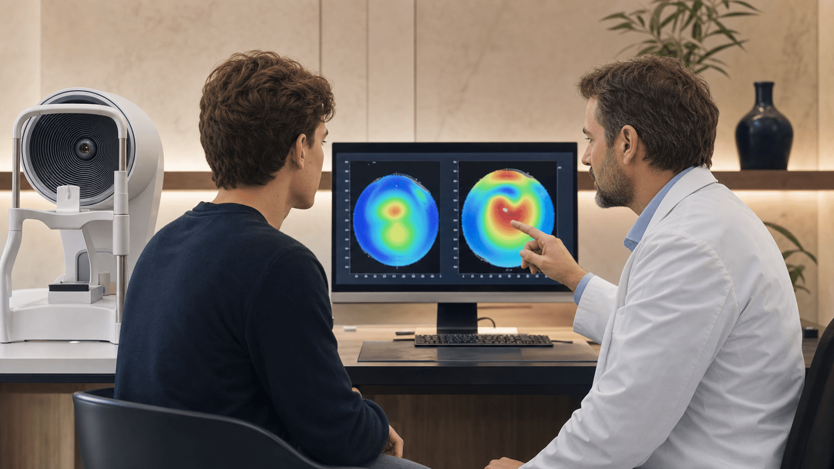
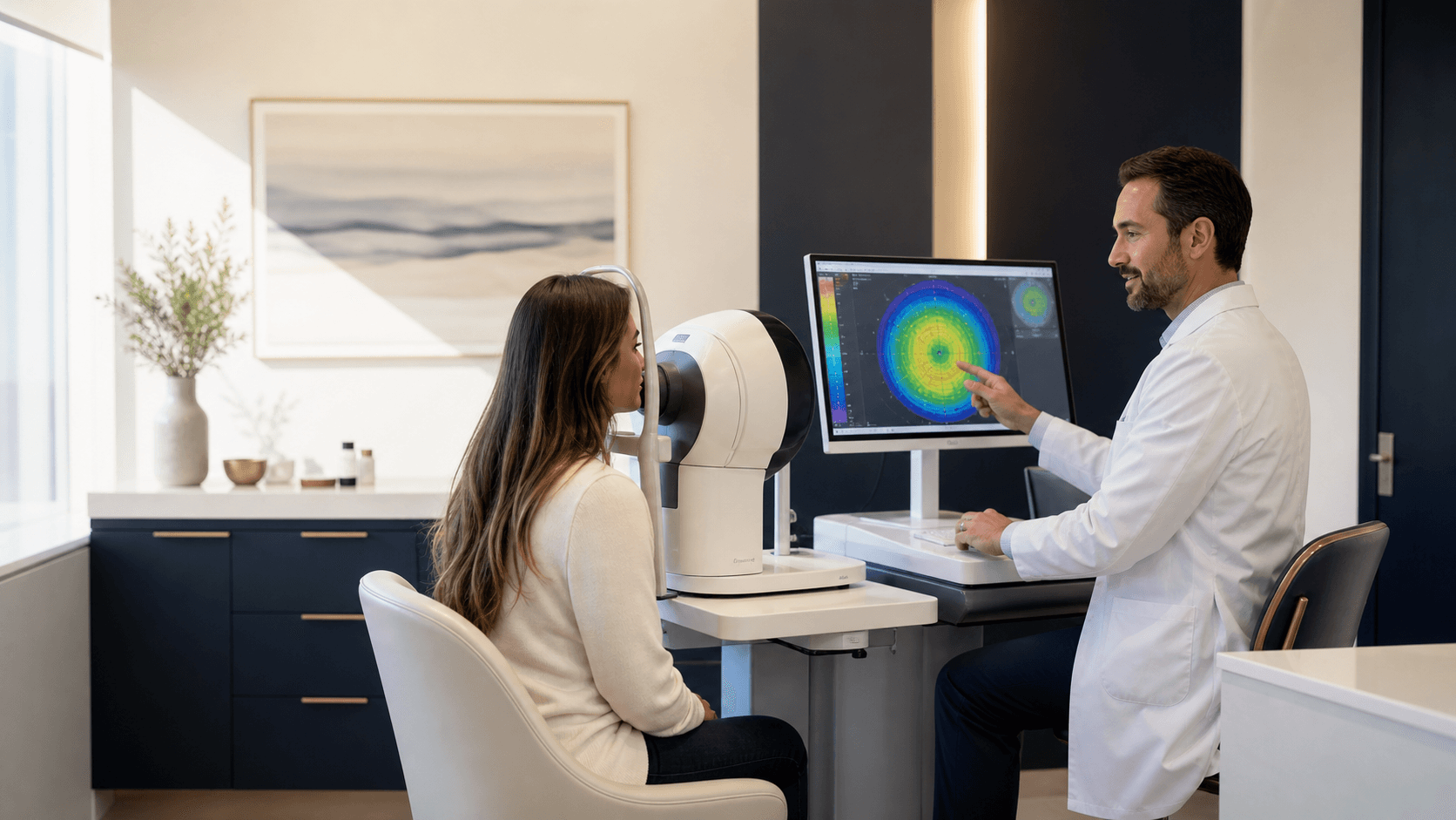
A cornea evaluation may use topography, tomography, pachymetry, refraction history, and prior maps to decide whether the shape is stable or progressing.
That distinction matters because:
- Glasses or specialty contact lenses can improve vision but do not stop documented progression.
- Corneal cross-linking is intended to strengthen a progressing cornea.
- CTAK or other reshaping strategies are considered only in selected eyes and do not replace the stability question.
- Advanced scarring or anatomy may require transplant-level consultation.
Start with the keratoconus condition guide if the diagnosis is already known.
Corneal scarring, haze, or an old injury
Scarring can follow infection, trauma, inflammation, prior surgery, or another corneal disorder. The effect on vision depends on the scar's depth, location, size, and whether the corneal shape is distorted.
A cornea-focused examination can help answer:
- Is the scar active or stable?
- Is vision limited by opacity, irregular astigmatism, or both?
- Would glasses, specialty lenses, surface treatment, or referral for surgery be appropriate?
- Is another part of the eye also limiting vision?
Treatment should follow that diagnosis. Not every visible scar needs surgery, and not every blurred eye can be improved by treating the cornea alone.
Contact lenses no longer fit or vision is unstable
Rigid and scleral lenses can provide excellent vision for irregular corneas, but poor tolerance can develop from dryness, fit, progression, inflammation, or scarring.
Bring the lens parameters, fit notes, and prior maps if possible. A cornea evaluation can separate a lens-fit problem from a changing cornea and determine whether the next step is a new lens design, surface treatment, cross-linking assessment, or surgical referral.
A pterygium is growing or changing vision
A pterygium is a growth of conjunctival tissue onto the cornea. It may remain small, or it may cause redness, irritation, induced astigmatism, and visual distortion as it extends toward the center.
Surgery is not automatically required because a pterygium exists. Evaluation may include slit-lamp examination, photographs, refraction, and corneal topography when the shape is affected.
The pterygium surgery page explains when growth, symptoms, vision, and recurrence planning enter the decision.
Severe dry eye or surface disease is affecting vision
Dry eye is not always only a tear problem. Meibomian gland dysfunction, blepharitis, Demodex, ocular rosacea, allergy, medication effects, autoimmune disease, exposure, and corneal nerve changes can overlap.
A cornea or ocular-surface evaluation is useful when:
- Vision fluctuates with blinking.
- Artificial tears are not enough.
- There is persistent staining or recurrent erosion.
- Contact lenses have become intolerable.
- Symptoms are delaying LASIK, PRK, cataract surgery, or lens replacement.
- There is concern for scarring, infection, or another surface condition.
Use the dry eye clinic page for a cause-based treatment workup.
Recurrent corneal erosion or repeated morning pain
Some patients wake with sharp pain, tearing, and light sensitivity because the corneal surface epithelium does not adhere normally. Prior abrasion, epithelial basement membrane changes, dry eye, or another surface disorder may contribute.
Repeated episodes deserve an examination. The plan can range from lubrication and surface protection to prescription treatment or a procedure, depending on the cause and severity.
A second opinion before LASIK, PRK, CXL, or another surgery
Corneal shape and thickness affect refractive-surgery safety. A second opinion may be appropriate when:
- You were told the cornea is thin or irregular.
- One practice recommends LASIK and another recommends PRK or EVO ICL.
- Topography suggests early keratoconus or ectasia risk.
- Prior LASIK or PRK vision is changing.
- CXL was recommended without a clear explanation of progression.
- You are comparing CXL, specialty lenses, CTAK, or transplant-level care.
Bring prior maps. A single scan cannot show progression unless it can be compared with a reliable earlier measurement.
What happens during a cornea-focused evaluation?
The visit may include:
- Visual acuity and refraction.
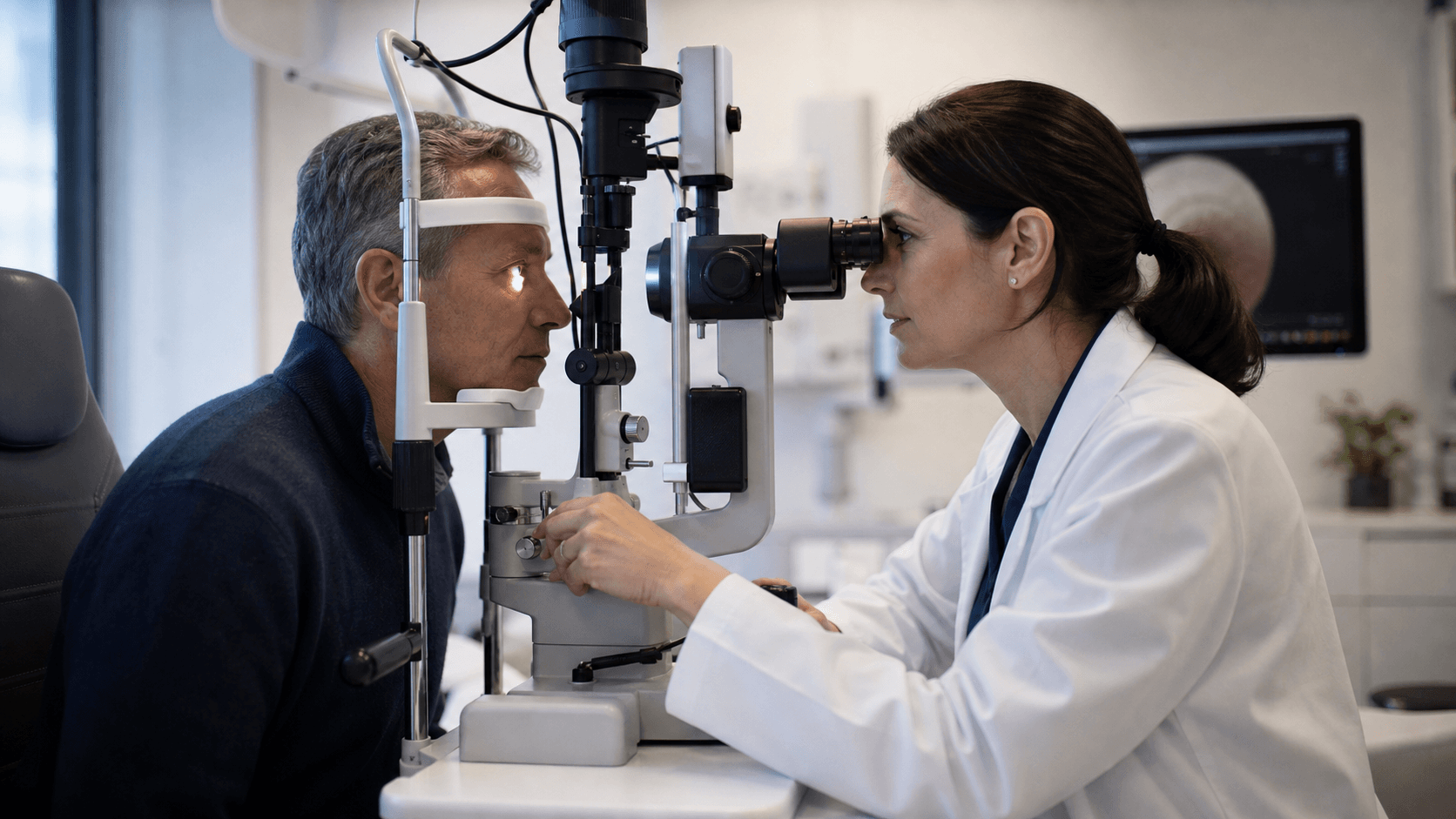
- Slit-lamp examination.
- Corneal topography and tomography.
- Pachymetry, or corneal thickness measurement.
- Fluorescein staining and tear-film review.
- Eyelid and meibomian gland examination.
- Review of contact lenses and prior procedures.
- Dilated examination when the symptoms or surgical decision require it.
Not every patient needs every test. The workup should end with a clear next step: monitor, treat the surface, repeat measurements, fit a specialty lens, consider CXL or pterygium surgery, or refer for a level of care the practice does not provide.
Cornea specialist versus ophthalmologist
An ophthalmologist is a medical doctor who diagnoses eye disease and can perform eye surgery. A cornea specialist is an ophthalmologist with additional focus or fellowship training in corneal and external disease.
The most important practical question is whether the clinician can perform the needed diagnostic workup and recognize when subspecialty referral is safer. A practice should not force every corneal problem into a procedure it happens to offer.
Bring useful records to the visit
When available, bring:
- Prior topography or tomography.
- Glasses prescriptions showing change over time.
- Contact-lens records.
- LASIK, PRK, CXL, or transplant operative notes.
- Medication and allergy lists.
- Photographs that show intermittent redness or swelling.
- Records from urgent-care or emergency visits.
Start with the cornea specialist page, compare cornea and dry-eye procedures, or schedule an evaluation at Visionary Eye Surgery in Plano.
Sources
Medically Reviewed by Dr. Shehz, DO
Dr. Shehzad Batliwala, DO—better known as Dr. Shehz—is a board-certified ophthalmologist and eye surgeon who brings both technical precision and genuine compassion to every patient he treats.
Explore this content with AI
Open this article in your preferred AI assistant for a plain-English summary, action steps, and questions to ask before booking a consultation.
Want a direct answer about your eyes? A complimentary consultation gives you a clear, written recommendation.