A pterygium can come back after surgery.
That is the concern most patients do not hear enough about before the first removal.
The goal of pterygium surgery is not only to remove the visible growth. The goal is to remove it in a way that protects comfort, corneal shape, appearance, and recurrence risk over time.
Why pterygium can recur
Pterygium is not just a surface bump. It is fibrovascular tissue that grows from the conjunctiva toward or onto the cornea, often influenced by UV exposure, dryness, irritation, inflammation, wind, dust, and individual healing tendency.
After removal, the treated area has to heal. If the surface remains inflamed, exposed, or unstable, tissue can grow back across the limbus and onto the cornea.
Recurrence risk can be affected by:
- The surgical technique used.
- Whether bare sclera was left exposed.
- Whether a graft was used.
- Prior pterygium surgery.
- Younger age.
- Larger or more inflamed pterygium.
- Ongoing UV exposure.
- Dry eye or ocular surface inflammation.
- Incomplete follow-up or drop use after surgery.
That is why recurrence prevention starts before surgery, not after the growth comes back.
Why older bare-sclera removal is different
Older pterygium surgery often focused on removing the growth and leaving the sclera exposed.
That approach is called bare-sclera excision. It can remove the pterygium, but the uncovered area creates a higher risk that fibrovascular tissue will regrow.
Modern pterygium surgery usually closes the area with tissue. The most common recurrence-focused repair is conjunctival autograft.
What conjunctival autograft means
Conjunctival autograft means your surgeon removes the pterygium, then moves a small piece of healthy conjunctival tissue from your own eye into the treated area.
The graft covers the bare surface, supports healing, and creates a healthier barrier at the limbus. Published ophthalmology literature generally supports conjunctival autograft as a lower-recurrence approach compared with leaving bare sclera exposed.
At Visionary Eye, pterygium surgery planning includes whether conjunctival autograft repair is appropriate for the growth pattern, ocular surface, prior surgery history, and future eye-care needs.
Autograft vs amniotic membrane vs bare sclera
Not all pterygium surgery is the same.
Bare-sclera removal is the older approach: remove the growth and leave the treated surface uncovered. It can look simple, but recurrence risk is much higher because the healing surface is exposed.
Conjunctival autograft repair covers the treated area with healthy tissue from the patient's own eye. Published ophthalmology literature generally supports conjunctival autograft as a lower-recurrence option than bare sclera, and many studies show it compares favorably with amniotic membrane for recurrence prevention.
Amniotic membrane can still be useful in selected cases. It may be considered when the defect is large, when the surgeon wants to preserve conjunctiva for future glaucoma surgery, when scarring limits available tissue, or when ocular-surface reconstruction needs a different approach.
Mitomycin C or 5-fluorouracil may be discussed in some recurrent or high-risk cases, but those medications are not automatic add-ons. They can carry their own risks, so the safer conversation is individualized: primary vs recurrent pterygium, amount of inflammation, tissue quality, glaucoma history, and long-term surface health.
For recurrent pterygium, the important question is not just who can remove it. It is which repair strategy protects the cornea and conjunctiva over time.
What if my pterygium already came back?
A recurrent pterygium is usually more complex than a first-time pterygium.
There may be scar tissue. The conjunctiva may be less mobile. The cornea may already have irregular astigmatism. The growth may be more inflamed or more adherent than before.
A recurrence-focused evaluation should check:
- Where the prior surgery was done.
- Which technique was used.
- How quickly the growth returned.
- Whether a graft was used.
- Whether mitomycin, amniotic membrane, glue, or sutures were part of the plan.
- Whether the pterygium is changing corneal shape.
- Whether the eye has dry eye, blepharitis, or other surface inflammation.
- Whether glaucoma surgery may ever require conjunctival tissue.
Bring prior records if you have them. They can help the surgeon understand what tissue was used and what should be preserved.
How pterygium recurrence can affect vision
Recurrence is not only cosmetic.
A returning pterygium can pull on the cornea and change its shape. That can create or worsen astigmatism, blur, glare, ghosting, and contact lens intolerance.
If the growth approaches the visual axis, the vision conversation becomes more urgent. If it remains small but inflamed, the first step may be surface control and monitoring.
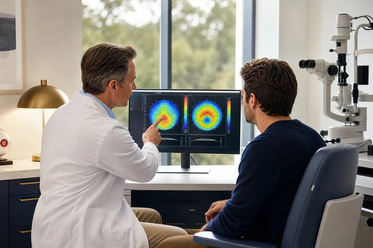
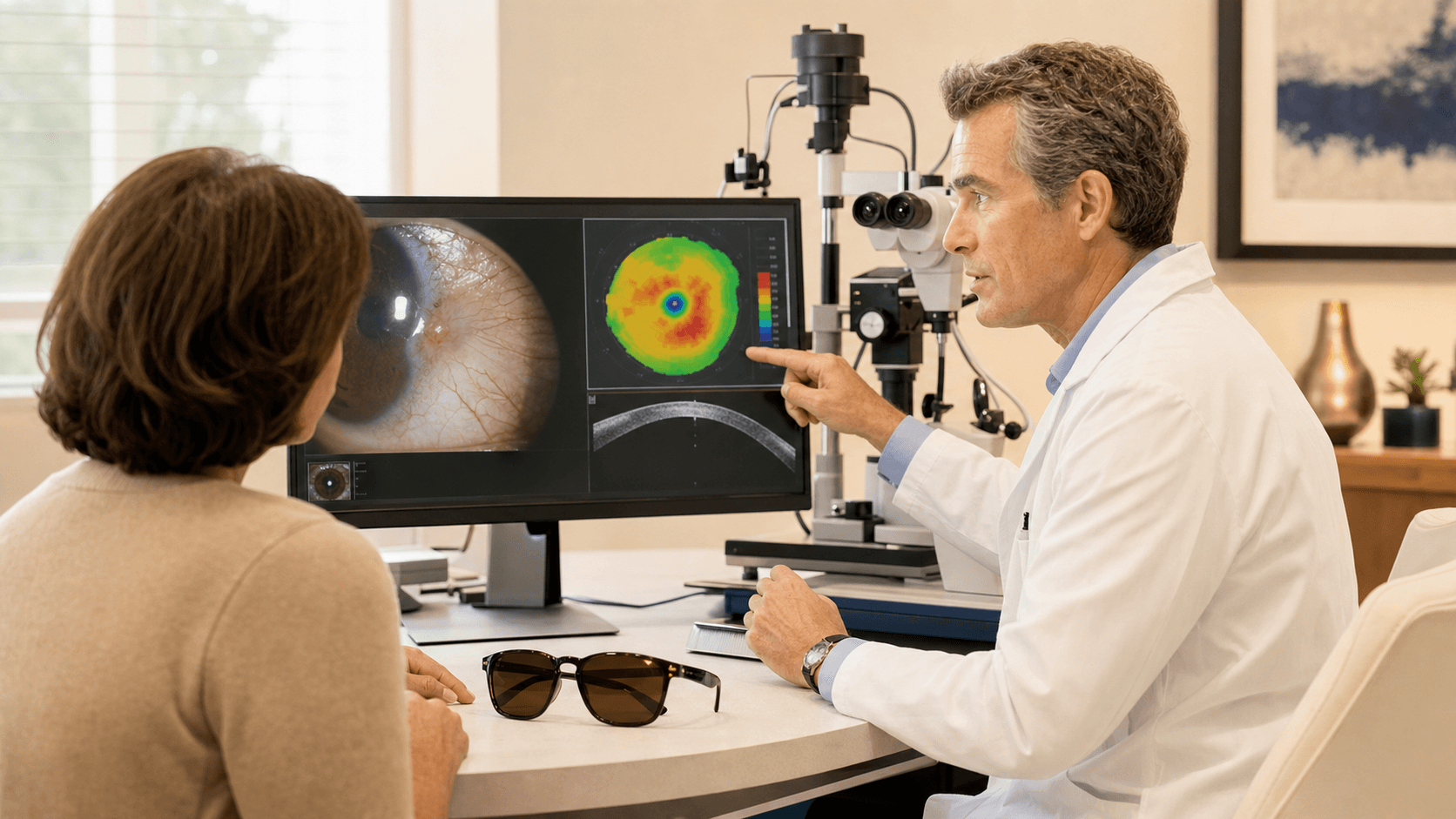
That distinction is why a pterygium surgeon should measure the growth and the corneal shape rather than deciding from appearance alone.
Recovery and prevention after surgery
Patients can help reduce recurrence risk by following the postoperative plan closely.
Common instructions may include:
- Using prescribed anti-inflammatory and antibiotic drops.
- Attending early follow-up visits.
- Avoiding eye rubbing.
- Wearing UV-blocking sunglasses outdoors.
- Treating dry eye or eyelid inflammation when present.
- Avoiding dusty or windy exposure during early healing when possible.
Redness, tearing, foreign-body sensation, and light sensitivity can happen during early healing. The graft can look more noticeable before it settles. Your surgeon should explain what is expected and what deserves a call.
Why the first year matters
Recurrence often declares itself during the early healing window and the months after surgery, which is why follow-up is part of the procedure rather than an optional extra.
Call the office if you notice a new fleshy edge moving toward the cornea, worsening redness after initial improvement, new pulling or restriction, worsening blur, or symptoms that feel similar to the original growth. Early inflammation control and measurement can help the surgeon decide whether the eye is healing normally or needs a change in treatment.
Sources and clinical references
- Pterygium clinical overview - American Academy of Ophthalmology EyeWiki
- Eye health and surgery patient guide - American Academy of Ophthalmology
These sources support general clinical context. Recurrence risk and repair strategy depend on the individual eye and prior surgery.
Related resources
Bottom line
Pterygium can recur after surgery, especially when the surface is left exposed, inflammation is not controlled, UV exposure continues, or the case is already recurrent.
Conjunctival autograft repair is designed to lower recurrence risk compared with older bare-sclera approaches, but no surgery can promise that a pterygium will never return.
The right plan starts with confirming the diagnosis, measuring corneal involvement, reviewing prior surgery if any, and choosing a repair strategy that respects both recurrence risk and the rest of the eye.
Start with Pterygium Surgery in Plano, read Do I Need Pterygium Surgery?, or schedule a pterygium surgeon evaluation if the growth is changing or has returned.
Your exam can clarify which options fit your eyes. Visit our Plano team from anywhere in DFW.
Book a free visit